![]()
![]()
Syed Saleem MujtabaI; Simon LedinghamI; Asif Raza ShahI; Stephan SchuelerI; Stephen ClarkI; Thasee PillayI
DOI: 10.21470/1678-9741-2017-0157
AVR = Aortic valve replacement
CPB = Cardiopulmonary bypass
ECC = Extracorporeal circulation
POD = Postoperative day
TAVI = Transcatheter aortic valve implantation
INTRODUCTION
Surgical aortic valve replacement (AVR) still represents the gold standard in patients with severe aortic valve stenosis[1]. Owing to increasing age of the patient population in the Western world, there has been an increase in the prevalence of patients with valvular heart disease eligible for AVR[2]. Given the increasing number of co-morbidities and the increasing age of patients, a tendency has emerged to use biological valve implants thus avoiding the need for long-term anticoagulation therapy. Although the concept of transcatheter aortic valve implantation (TAVI) appears attractive, the calcified aortic valve is not removed during this procedure. Therefore, paravalvular leakage remains an important issue with this technique[3]. Other important concerns are access site-related problems and device malpositioning.
The recent introduction of sutureless bioprostheses may offer an additional tool in the therapeutic armamentarium as these valves do not need to be sutured into place resulting in shorter cross-clamp and cardiopulmonary bypass (CPB) durations which may be beneficial in older patients with co-morbid conditions. Moreover, due to the absence of a sewing ring, these valves exhibit favourable hemodynamic properties. Excellent outcomes have been demonstrated with sutureless AVR in minimally invasive surgical setting[4].
We noticed that sutureless valves are associated with greater drops in postoperative platelet counts compared to sutured valves. Albacker[5] reported this phenomenon with sutureless AVR and the aim of this study was to investigate this observation and try to find an explanation for its occurrence.
METHODS
Patients
It is a retrospective, observational study. All the patients who underwent isolated sutureless Perceval AVR between June 2014 and November 2016 were compared with patients who had isolated AVR with Carpentier-Edwards Perimount Magna Ease valve between June 2015 and January 2017. Any non-isolated AVR cases i.e.: coronary artery bypass grafting, mitral and tricuspid valve surgery, myomectomy, ascending aorta surgery were excluded. Moreover, all the redo, emergency and infective endocarditis patients were excluded.
All the sutureless AVRs were performed by the six different surgeons in our institution during that period. Data was collected on all the patients who underwent sutured AVR by the same surgeons during the same time period (101 patients) as a control group.
A total of 72 patients received isolated Perceval bioprosthesis and 101 patients had Perimount Magna Ease valve. The Percevel S valve sizes were small, medium, large and extra-large. For statistical purposes they were taken as equivalent to 19-21, 23, 25 and 27 mm Perimount Magna Ease sizes, respectively.
Severe thrombocytopenia was defined as a platelet count <50x109/Lit during the postoperative period. The platelet count was determined preoperatively, day of surgery and every day until day 5 in the postoperative period. In-hospital mortality was defined as any death occurring within first 30 days after surgery. Aspirin and clopidogrel was discontinued 7 days before surgery and warfarin was stopped 5 days before operation.
Surgical Approach
A complete median sternotomy was performed and extracorporeal circulation (ECC) with moderate hypothermia (34°C) was employed in every patient. Antegrade cold blood cardioplegia was administered after application of cross clamp. The choice of prosthesis implanted was made by the surgeon. Perioperative transesophageal echocardiogram was performed routinely to confirm correct position of the valve, exclude any paravalvular leak and evaluate valve hemodynamics.
The Perimount Magna Ease valve was rinsed with normal saline for 3 minutes before implantation while Perceval valve does not require any rinsing before implantation. Perimount Magna Ease valves were intra implanted with interrupted or semi continuous 2/0 prolene sutures.
The Perceval valve is a surgical bioprosthetic heart valve consists of glutaraldehyde-fixed bovine pericardium treated with homocysteic acid in order to remove the free aldehyde residues and prevent the calcification process. It is fixed in a metal cage made up of an alloy of nickel and titanium, known as nitinol.
Three 4/0 polypropylene guiding sutures were passed at the nadir of the aortic annulus. An appropriately sized prosthesis was collapsed in a side table and placed into the manufacturer's holder. The three guiding sutures were passed through the three green holes arising from the annular ring of the prosthesis, which was consequently seated on the debrided annulus. Once the delivery system is in position, the stent is deployed by turning the release screw and leaving the valve in place. The delivery system and the guiding sutures are removed. The field was rinsed with warm saline, and the prosthesis was dilated at four atmospheres for 30 seconds.
On first postoperative day, patients were started on aspirin 150 mg orally and low molecular weight heparin subcutaneously for deep vein thrombosis prophylaxis.
Statistical Analysis
All data has been extracted from the Dendrite PATS CS2010F SCTS v4.1.2 database. Date of data extract was 25.07.17 to cover the period from 01.06.2014 to 31.01.17. Analysis was performed using MS Excel. Numerical values were compared using an independent t-Test, with a two-tailed distribution assuming unequal variances. Categorical variables were compared using Chi Square χ2 analysis.
RESULTS
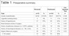
The preoperative characteristics of patients are depicted in Tables 1 and 2. There was no difference between Group A and B regarding history of preoperative renal impairment or pulmonary disease. Group A has more female patients (A=52% vs. B=31%; P≤0.004), patients with hypertension (79% vs. 60%; P=0.01), neurological dysfunction (17% vs. 7%; P=0.05) and angina (54% vs. 37%; P=0.03). In group A, patients were older (range: 54-84, mean 74 vs. 47-86, mean 70 vs. range 34-91, mean 71; P=0.001) and with higher logistic EuroSCORE (3.07 vs. 1.87; P=0.001).
| Perceval | Perimount | Chi-squared Test | |||
|---|---|---|---|---|---|
| Total | n=72 | % | n=101 | % | P |
| Gender (male/female) | 34/38 | 47/52% | 69/31 | 69/31% | 0.004 |
| Cigarette smoking history | 3 | 4% | 11 | 11% | 0.106 |
| History of hypertension | 57 | 79% | 60 | 60% | 0.01 |
| Renal disease at time of surgery | __ | __ | 1 | 1% | 0.40 |
| History of pulmonary disease (i.e: COPD, asthma) | 16 | 22% | 19 | 19% | 0.61 |
| History of neurological disease (i.e.: TIA, CVA) | 12 | 17% | 7 | 7% | 0.05 |
| Angina Status pre-surgery | 39 | 54% | 37 | 37% | 0.03 |
| 0. No angina | 33 | 46% | 63 | 88% | |
| 1. No limitation of physical activity | 16 | 22% | 18 | 25% | |
| 2. Slight limitation of ordinary activity | 11 | 15% | 11 | 15% | |
| 3. Marked limitation of ordinary physical activity | 10 | 14% | 6 | 8% | |
| 4. Symptoms at rest or minimal activity | 2 | 3% | 2 | 3% | |
| Dyspnoea status pre-surgery | 65 | 90% | 82 | 82% | 0.13 |
| 1. No limitation of physical activity | 7 | 10% | 18 | 18% | |
| 2. Slight limitation of ordinary physical activity | 23 | 32% | 39 | 39% | |
| 3. Marked limitation of ordinary physical activity | 41 | 57% | 40 | 40% | |
| 4. Symptoms at rest or minimal activity | 1 | 1% | 3 | 3% | |
| History of diabetes mellitus | 13 | 18% | 20 | 20% | 0.75 |
| Preoperative heart rhythm | |||||
| 0. Sinus rhythm | 55 | 76% | 84 | 87% | |
| 1. Atrial fibrillation/flutter | 16 | 22% | 12 | 12% | 0.07 |
| 2. Complete heart block/pacing | 1 | 1% | __ | __ | 0.24 |
| 3. Other abnormal rhythm | __ | __ | 1 | 1.0% | |
| Ejection fraction category | |||||
| 1. Good (LVEF > 50%) | 58 | 81% | 84 | 84% | |
| 2. Fair (LVEF 30-50%) | 12 | 17% | 15 | 15% | 0.77 |
| 3. Poor (LVEF < 30%) | 2 | 3% | 1 | 1% | 0.38 |
| Perceval | Perimount | t-Test | |||||
|---|---|---|---|---|---|---|---|
| Range | Mean | Median | Range | Mean | Median | Two-Sample Assuming Unequal Variances | |
| Age of patients at time of procedure | 54-84 | 74 | 75 | 47-86 | 70 | 70.5 | <0.001 |
| Logistic EuroSCORE comparison | 0.53-18.89 | 3.07 | 2.41 | 0-7.57 | 1.87 | 1.44 | 0.001 |
| Height (cm) | 140-185 | 164.1 | 163 | 138-185 | 167.3 | 169 | 0.029 |
| Weight (kg) | 40.3-158 | 82.2 | 80.1 | 50-181.6 | 84.90 | 81.1 | 0.387 |

Greater number of L and XL size valves was implanted in Perceval valve group, and of 23 and 25 mm prosthesis in Perimount Magna Ease group (Table 3).
| Perceval valve sizes | N | % | Perimount Magna valve sizes | N | % |
|---|---|---|---|---|---|
| 19 | 1 | 1 | |||
| Small | 8 | 11 | 21 | 20 | 11 |
| Medium | 20 | 28 | 23 | 42 | 42 |
| Large | 22 | 31 | 25 | 29 | 29 |
| X large | 22 | 31 | 27 | 10 | 18 |
| 29 | __ | __ | |||
| Total | 72 | 101 |

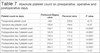
When Perceval valve sizes S, M, L and XL were compared with their counterpart Perimount Magna Ease valve sizes, there was more significant thrombocytopenia in Perceval group (Table 4). Haematology data is depicted in Tables 5, 6 and 7. Among the Perceval valve group, 5 (6%) patients had severe thrombocytopenia. Their peak reduction was on day 3 with 58% of their preoperative platelet value. While in Perimount Magna group none of them had severe thrombocytopenia. Their peak reduction was on day 2 with 44% of preoperative value which is significantly less then Perceval valve group. Perceval valve group required more blood (1.5 vs. 0.7 units) (P=0.009) and platelet (0.3 vs. 0.1 platelets pools; P=0.007) transfusion. No significant difference in absolute platelet count between two groups on preoperative and operation days was observed. Platelet count was significantly reduced in Perceval group from day 1-6 and 20% patients came back to preoperative level at discharge. Forty-four percent of Perimount patients came back to preoperative value at the time of discharge. Forty-one percent of Perimount group had moderate thrombocytopenia while this value was 26% in Perceval valve group.
| Perceval valve Size % | Perimount Magna valve Size % | P value |
|---|---|---|
| Small 66.11% | 19-21 mm 48.36% | 0.0001 |
| Medium 56.28% | 23 mm 43.25% | 0.0003 |
| Large 59.41% | 25 mm 44.85% | 0.0003 |
| X large 56.89% | 27-29 mm 39.3% | 0.0005 |

| Perceval valve N=72 |
Perimount Magna N=101 |
P | |
|---|---|---|---|
| Maximum drop in platelet count: "which day" | Mean 2.3 Median 2 Range 0-5 |
Mean 1.7 Median 2 Range 0-6 |
0.0005 |
| Maximum drop in platelet count: "what %" | Mean 58.0% Median 58.3% Range 35-84% |
Mean 44.3% Median 45.0% Range 14-64% |
<0.0001 |
| Blood transfusion: units | Mean 1.5 Median 0.5 Range 0-10 |
Mean 0.7 Median 0 Range 0-8 |
0.0091 |
| Platelet transfusion: pool of platelets | Mean 0.3 Median 0 Range 0-4 |
Mean 0.1 Median 0 Range 0-2 |
0.0075 |
| FFP Transfusion: units | Mean 0.2 Median 0 Range 0-4 |
Mean 0.1 Median 0 Range 0-3 |
0.21 |

| Patients having postoperative thrombocytopenia | Perimount Magna valve | Perceval valve | P value |
|---|---|---|---|
| Moderate thrombocytopenia: <100 x 109 | N=26 (26%) | N=34 (41%) | 0.008 |
| Severe thrombocytopenia: <50 x 109 | __ | N=5 (6%) | |
| Patients whose platelet level returned to preoperative level following surgery/did not return to preoperative | Perimount Magna valve N=44/57 (44/56%) |
Perceval valve N=20/56 (26/74%) |
0.018 |

| Platelet count on days | Perimount Magna valve | Perceval valve | P value |
|---|---|---|---|
| Preoperative platelet count | 244 | 239.8 | =0.73 |
| Operation day platelet count | 158.6 | 144.6 | =0.07 |
| Postoperative day 1 platelet count | 158.7 | 132.8 | =0.0007 |
| Postoperative day 2 platelet count | 145.9 | 112.8 | <0.0001 |
| Postoperative day 3 platelet count | 158.4 | 111.4 | <0.0001 |
| Postoperative day 4 platelet count | 183.5 | 130.0 | <0.0001 |
| Postoperative day 5 platelet count | 216.4 | 157.5 | <0.0001 |
| Postoperative day 6 platelet count | 260.3 | 174.4 | <0.0001 |
| Postoperative day 7 platelet count | 254.6 | 205.7 | =0.23 |
Intraoperative and early postoperative variables are summarized in Tables 8 and 9. Cross clamp (range 23-76 min, mean 39 min vs. 24-133 min, mean 54 min; P=0.001)) and bypass time (range 25-119 min, mean 59 min vs. range 19-175 min, mean 71 min; P=0.001) were shorter in isolated Perceval valve group (Group A1) compared to isolated conventional valve group (Group B1).
| Perceval | Perimount | t-Test: Two-Sample Assuming Unequal Variances | |||||
|---|---|---|---|---|---|---|---|
| Range | Mean | Median | Range | Mean | Median | P | |
| Cumulative cross-clamp time (min) | 23-76 | 39.2 | 38 | 24-133 | 54.1 | 48.5 | <0.001 |
| Cumulative bypass time | 25-119 | 59.3 | 56.5 | 19-175 | 71.6 | 63.5 | <0.001 |
| Post operative blood loss @ 12 hours | 15-2000 | 304 | 212.5 | 100-2000 | 359.7 | 300 | 0.197 |
| ITU stay in days | 1-13 | 3.2 | 2 | 1-17 | 1.9 | 1 | 0.005 |

| Perceval | Perimount | Chi-squared Test | |||
|---|---|---|---|---|---|
| Total | n=72 | % | n=100 | % | P |
| Reoperation for bleeding, tamponade | 2 | 2.78% | 2 | 2.00% | 0.738 |
| Patient status at discharge (mortality) | 1 | 1.39% | __ | 0.00% | 0.237 |

Perceval valve patients spent more time in ICU compared to Perimount Magna patients (3.2 days vs. 1.9 days; P=0.005). No significant difference in postoperative neurological dysfunction, renal impairment, atrial fibrillation, permanent pacemaker requirement or mortality could be observed.
DISCUSSION
Our retrospective study showed that there is a relationship between Perceval S valve implantation and severe thrombocytopenia. Severe thrombocytopenia in our study was not associated with higher mortality and morbidity, and none of these deaths was related to severe thrombocytopenia.
Sánchez et al.[6] compared the incidence of thrombocytopenia after AVR with Perceval S Sutureless Bioprosthesis (n=27) and Mitroflow prostheses (n=50). The incidence of severe thrombocytopenia was significantly higher (P=0.046) in Perceval S patients than in Mitroflow patients. The platelet count recovered in all patients with severe thrombocytopenia.
The biological structure of the Perceval S valve and Freedom Solo stentless bioprosthesis are very similar[6].
Hilker et al.[7] compared the postoperative courses of platelet counts in patients having had AVR with stentless prostheses (Sorin Biomedica Freedom Solo [SOLO]) or stented prostheses (Carpentier Edwards Perimount [PM]). A higher occurrence of platelet levels below 100 Gpt/l between the second and the fifth postoperative day (POD) was found in the SOLO-group (71.9%) compared with the other biological substitute PM (36.6%).
Miceli et al.[8] evaluated the postoperative evolution of platelet count and function after AVR in 116 patients undergoing isolated stentless biological AVR with Freedom Solo and compared with 206 patients who received stented biological valves. Freedom Solo implantation was associated with a higher incidence of thrombocytopenia compared with the control group (24.1% vs. 4.4%, P<0.0001).
Piccardo et al.[9] also studied the incidence and clinical impact of thrombocytopenia in patients receiving AVR with Freedom Solo bioprosthesis and Carpentier-Edwards Perimount pericardial prosthesis. They reported that severe thrombocytopenia occurred in 25% and 3% of patients with Freedom Solo and Perimount bioprostheses, respectively (P<0.0001).
Yerebakan et al.[10] compared platelet counts within 2 weeks after implantation of either a stentless (Sorin Freedom Solo) or a stented (Sorin Mitroflow) bovine pericardial bioprosthesis. In the Mitroflow group, the mean platelet count moderately dropped to a minimum of 60% of the initial value on 3rd POD and fully recovered on 8th POD. In the Freedom Solo group, platelet loss was significantly more severe (minimum relative value 25% on 4th POD) with no recovery during follow-up. However, there were no other complications reported in association with thrombocytopenia related to the use of these Freedom Solo valves and the phenomenon was transient and resolved without clinical consequences or hemodynamic dysfunction[9,10].
Platelet count decrease has also been reported after percutaneous coronary intervention[11]. Gallet et al.[12] studied the effect of transcatheter (via femoral artery) aortic valve implantation (TAVI) on platelet count. They observed that platelet count systematically decreased after TAVI, with an average decrease of 34±15%, and a decrease in platelet count reported to be associated with in-hospital major adverse cardiovascular events and strongly influenced patient outcome. In the setting of TAVI procedures platelet activation can be caused by endothelial damage caused by prosthesis implantation, fibrinogen binding on metallic armatures, and shear stress modifications due to prosthesis implantation[13].
Van Straten et al.[14] compared thrombocytopenia after AVR with mechanical and biological valves. They concluded that patients undergoing AVR with the Carpentier-Edwards Perimount bioprosthesis or a Medtronic Freestyle stentless bioprosthesis had a lower minimum platelet count within the first five POD, compared to patients receiving ATS and St. Jude Medical mechanical prostheses.
In our study, the drop-in platelet count started to occur during the 3rd POD with very slow recovery toward the 7th to 10th POD. The lowest drop in platelet count was down to 22% of the preoperative baseline level and occurred in patients implanted with the Perceval valve. As shown in other studies, there were no other clinical consequences associated with thrombocytopenia that was transient but resulted in more transfusion requirements.
The bovine pericardium leaflets of both Perceval S valve and Mitroflow bioprosthesis are fixed in a process using gluteraldehyde. The Mitroflow prosthesis is stored in a solution of gluteraldehyde and needs rinsing before it is used. On the other side, the Perceval S prosthesis and Freedom Solo prosthesis are detoxified with homocysteic acid to eliminate any residual aldehyde and then stored in an aldehyde free solution; so, it does not require rinsing prior to use. Homocysteic acid can have a damaging effect on vessel endothelial cells and can precipitate platelet aggregation resulting in both thrombocytopenia and thrombotic complications[15]. These valves are carefully washed after detoxification with a homocysteic acid-free solution and then stored in jars filled with a homocysteic acid-free storage solution. Consequently, the residual amount of homocysteic acid, which could be transferred to the patient during the valve implantation, is negligible and the resulting concentration in blood is extremely low, so cannot be blamed for the postoperative thrombocytopenia. Moreover, the same solution is used for other bioprosthesis[16], without causing higher incidence of thrombocytopenia.
Mechanical stress and hemodynamic turbulence have also been proposed as a cause of thrombocytopenia after Freedom Solo prosthesis[10].
Small valve sizes have been described as causing some turbulence across the valve resulting in platelet activation or destruction, and consequently postoperative thrombocytopenia[7,9,14]. However, sutureless valves are well known for their superior hemodynamic performance and this hypothesis is quite unlikely.
Non-specific activation of platelets leading to diffuse consumption and thrombocytopenia after AVR with both mechanical and bioprosthetic aortic valves was suggested by Leguyader et al.[17]. Platelet activation could also result from valve manipulation or the presence of the metal stent.
The Perceval S valve has metal structure that serves as an anchoring system to the aortic valve, which could be the cause of turbulent flow, platelet rupture and mechanical structure. The metal stent may play a role in platelet activation and the possibility of paravalvular leaks may also trigger platelet activation and consumption. Moreover, the metal stents of these valves could interfere with the quality of images and could conceal or underestimate the degree of some paravalvular leaks[5].
Long CPB duration has been suggested by other studies to be associated with postoperative thrombocytopenia[14]. In our study the CPB durations were shorter in the sutureless group than the sutured one so it cannot be responsible for post-operative thrombocytopenia.
Severe thrombocytopenia is a common complication in patients who undergo cardiac surgery with ECC. It occurs as result of structural changes and activation of platelets, hemodilution, platelet aggregation and bleeding[18].
CONCLUSION
It seems to be an association between sutureless Perceval valve and severe thrombocytopenia, although it does not affect patient's mortality and morbidity. These patients had also higher rate of blood and platelets transfusion. A prospective randomised trial is needed to confirm our findings.
REFERENCES
1. Vahanian A, Alfieri O, Andreotti F, Antunes MJ, Barón-Esquivias G,Baumgartner H, et al. Guidelines on the management of valvular heart disease(version 2012): the Joint Task Force on the Management of Valvular Heart Diseaseof the European Society of Cardiology (ESC) and the European Association forCardio-Thoracic Surgery (EACTS). Eur J Cardiothorac Surg.2012;42(4):S1-44. [MedLine]
2. Bridgewater B, Gummert J, Kinsman R, Walton P. Towards globalbenchmarking: the Fourth EACTS Adult Cardiac Surgical Database Report.Oxfordshire: Dendrite Clinical Systems, Henley-on-Thames,;2010.
3. Martens S, Sadowski J, Eckstein FS, Bartus K, Kapelak B, Sievers HH,et al. Clinical experience with the ATS 3f Enable® suturelessbioprosthesis. Eur J Cardiothorac Surg. 2011;40(3):749-55.
4. Santarpino G, Pfeiffer S, Concistrè G, Fischlein T. Perceval Saortic valve implantation in mini-invasive surgery: the simple suturelesssolution. Interact Cardiovasc Thorac Surg. 2012;15(3):357-60.
5. Albacker TB. Thrombocytopenia associated with Perceval suturelessaortic valve replacement in elderly patients: a word of caution. Heart SurgForum. 2015;18(3):E093-7.
6. Sánchez E, Corrales JA, Fantidis P, Tarhini IS, Khan I, Pineda T, etal. Thrombocytopenia after aortic valve replacement with Perceval S suturelessbioprosthesis. J Heart Valve Dis. 2016;25(1):75-81. [MedLine]
7. Hilker L, Wodny M, Ginesta M, Wollert HG, Eckel L. Differences inthe recovery of platelet counts after biological aortic valve replacement.Interact Cardiovasc Thorac Surg. 2009;8(1):70-3.
8. Miceli A, Gilmanov D, Murzi M, Parri MS, Cerillo AG, Bevilacqua S,et al. Evaluation of platelet count after isolated biological aortic valvereplacement with Freedom Solo bioprosthesis. Eur J Cardiothorac Surg.2012;41(1):69-73. [MedLine]
9. Piccardo A, Rusinaru D, Petitprez B, Marticho P, Vaida I,Tribouilloy C, et al. Thrombocytopenia after aortic valve replacement withfreedom solo bioprosthesis: a propensity study. Ann Thorac Surg.2010;89(5):1425-30.
10. Yerebakan C, Kaminski A, Westphal B, Kundt G, Ugurlucan M, SteinhoffG, et al. Thrombocytopenia after aortic valve replacement with Freedom Solostentless bioprosthesis. Interact Cardiovasc Thorac Surg.2008;7(4):616-20.
11. De Labriolle A, Bonello L, Lemesle G, Roy P, Steinberg DH, Xue Z, etal. Decline in platelet count in patients treated by percutaneous coronaryintervention: definition, incidence, prognostic importance, and predictivefactors. Eur Heart J. 2010;31(9):1079-87.
12. Gallet R, Seemann A, Yamamoto M, Hayat D, Mouillet G, Monin JL, etal. Effect of transcatheter (via femoral artery) aortic valve implantation onthe platelet count and its consequences. Am J Cardiol.2013;111(11):1619-24.
13. Nobili M, Sheriff J, Morbiducci U, Redaelli A, Bluestein D. Plateletactivation due to hemodynamic shear stresses: damage accumulation model andcomparison to in vitro measurements. ASAIO J. 2008;54(1):64-72. [MedLine]
14. van Straten AH, Hamad MA, Berreklouw E, ter Woorst JF, Martens EJ,Tan ME. Thrombocytopenia after aortic valve replacement: comparison betweenmechanical and biological valves. J Heart Valve Dis.2010;19(3):394-9.
15. Piccardo A, Rusinaru D, Petitprez B, Marticho P, Vaida I,Tribouilloy C, et al. Thrombocytopenia after aortic valve replacement withfreedom solo bioprosthesis: a propensity study. Ann Thorac Surg.2010;89(5):1425-30.
16. Santarpino G, Pfeiffer S, Fischlein T. Thrombocytopenia afterfreedom solo: the mystery goes on. Ann Thorac Surg.2011;91(1):330.
17. Leguyader A, Watanabe R, Berbé J, Boumediene A, Cogné M, Laskar M.Platelet activation after aortic prosthetic valve surgery. Interact CardiovascSurg. 2006;5(1):60-4.
18. Weerasinghe A, Taylor KM. The platelet in cardiopulmonary bypass.Ann Thorac Surg. 1998;66(6):2145-52.
No financial support.
No conflict of interest
Authors' roles & responsibilities
SSM Drafting the work or revising it critically for important intellectual content; final approval of the version to be published
SL Drafting the work or revising it critically for important intellectual content; final approval of the version to be published
ARS Drafting the work or revising it critically for important intellectual content; final approval of the version to be published
SS Drafting the work or revising it critically for important intellectual content; final approval of the version to be published
SC Drafting the work or revising it critically for important intellectual content; final approval of the version to be published
TP Drafting the work or revising it critically for important intellectual content; final approval of the version to be published
Article receive on Monday, July 31, 2017
 All scientific articles published at rbccv.org are licensed under a Creative Commons license


All rights reserved 2017 / © 2024 Brazilian Society of Cardiovascular Surgery
DEVELOPMENT BY ![]()
 Read in English
 English PDF
 Print
Print
 Send this article by email
 How to cite this article
 Submit a comment
 Mendeley
 Pocket